Memorial Hermann Imaging Centers offer a full range of nuclear medicine studies conducted by certified professionals at convenient locations across the Greater Houston area.
If your doctor has ordered a nuclear medicine exam, you may be unsure of what to expect. To find out more about the procedure, contact us today and our experienced imaging center staff can answer your questions, verify your insurance coverage and schedule your appointment at a time and location that is convenient for you.
What is Nuclear Medicine?
Nuclear medicine is a branch of diagnostic imaging that examines the molecular processes in your body to help diagnose conditions like cancer or heart disease as well as disorders of the brain, intestines or endocrine system. By using trace amounts of a radioactive material, a special camera and a sophisticated computer system, nuclear medicine can provide critical information about how your body is functioning—helping you to avoid other forms of testing that might otherwise require surgery or other invasive procedures.
In some cases, nuclear medicine can even be used as targeted therapy to treat conditions such as an overactive thyroid, or certain cancers, like lymphoma.
How Does Nuclear Medicine Work?
During a nuclear medicine exam, you will be guided through a three-step process, which includes:
- Step One: A tiny amount of radioactive material, called a radiotracer or radiopharmaceutical, will be introduced into your body by way of intravenous injection (IV), inhalation of a gas or orally in capsule form. The chemical makeup of this radiopharmaceutical is specially designed based on the part of your body being scanned.
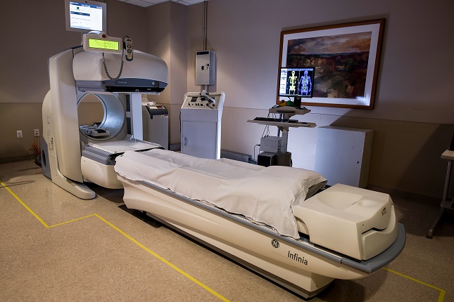
- Step Two: As the radioactive material travels through (or accumulates in) your body, it emits energy in the form of gamma radiation, which is detected by a special device called a gamma camera or scintillation camera. Unlike the imaging equipment for X-rays or computed tomography (CT), which both use ionizing radiation to scan the body, the gamma camera itself does not emit radiation. It only detects it. If nuclear medicine is being used to scan only a small section of your body, the nuclear technologist may opt to use a small handheld probe instead of the larger gamma camera.
- Step Three: Once this gamma radiation is detected, it is processed by a special computer using single-photon emission-computed tomography (SPECT) and converted into detailed, real-time images of the inside of your body. Because nuclear medicine can detect this radiation on the molecular level, it can provide comprehensive data about everything from blood flow and organ structure to your body’s metabolic processes.
Common Types of Nuclear Medicine Exams
Nuclear medicine can provide many diagnostic benefits depending on the part of your body being scanned. The following are a few of the most common ways nuclear medicine is used to diagnose injuries and disease:
- Heart: Imaging the heart was one of the original use cases for nuclear medicine, and it is just as important today as it was decades ago. Heart scans are used to monitor cardiac structure, function and blood flow, and to detect coronary artery disease and evaluate the need for bypass surgery or angioplasty.
- Lungs: Utilizing special equipment known as a xenon trap, a nuclear technologist can administer xenon gas into the mouth and nose, allowing the gamma camera to visualize respiratory function as the patient breathes. Blood flow to the lungs can also be evaluated.
- Bones: Though it is not the same as a standard X-ray, nuclear medicine is still a valuable diagnostic tool when checking bones for fractures, infection, tumors or metastatic disease.
- Brain: Nuclear medicine is an effective, low-radiation option to scan the brain for abnormalities including, but not limited to, Alzheimer’s and Parkinson’s disease, memory loss, seizures and tumors. If a patient is in a coma, nuclear medicine can help determine the extent to which the brain is functioning.
- Gallbladder: Using nuclear medicine, it is possible to visualize the intricate workings of the gastrointestinal (GI) tract in real-time. This can help doctors identify any inflammation or abnormal functions of the gallbladder and evaluate the post-operative effectiveness of gallbladder surgery.
- Thyroid: There are very few ways to measure thyroid function outside of nuclear medicine, and none are as effective. It is often used to aid in the diagnosis of blood cell disorders and hyperthyroidism.
- Kidneys: By injecting a radiopharmaceutical into the bloodstream, a nuclear technologist can use a gamma camera to visualize the blood supply and function of the kidneys, as well as the flow of urine from the kidneys and the presence of any urinary tract obstruction or reflux.
Nuclear medicine also can be an effective method for locating tumors and determining the spread (or staging) of certain cancers without the need for a biopsy. In this case, they are often paired with other imaging techniques such as X-rays or magnetic resonance imaging (MRI) in an effort to reach a conclusive diagnosis.
How is Nuclear Medicine Different from Other Imaging Procedures?
Nuclear medicine uses special gamma cameras to detect ionizing radiation coming from a radio drug that has been administered to a patient. By contrast, imaging procedures such as X-ray and computed tomography (CT) obtain images by beaming ionizing radiation through the body and capturing images.
Most imaging procedures produce static images, but images produced through nuclear medicine are in real time, much like an ultrasound. Nuclear medicine also differs in that it detects the presence of disease based on biological changes in tissue rather than changes in a patient’s anatomy.
Is Nuclear Medicine Safe?
According to the Society of Nuclear Medicine and Molecular Imaging (SNMMI), more than 20 million Americans receive nuclear medicine procedures each year to diagnose and treat a wide variety of conditions and diseases. While these procedures do expose patients to an extremely low dose of non-ionizing radiation, this risk is outweighed by the potentially lifesaving impact of the information they provide—information that would otherwise require exploratory surgery or other more costly and invasive procedures.
At Memorial Hermann, we are required to follow the standards for care set forth by the U.S. Food and Drug Administration’s Initiative to Reduce Unnecessary Radiation Exposure from Medical Imaging, which maintains that nuclear medicine exams must be performed with the lowest acceptable radiation levels for the shortest time necessary.
If you have any questions or concerns about the safety or necessity of a particular nuclear medicine exam, contact your physician or a Memorial Hermann Imaging Center staff member. We will be more than happy to assist you.
Possible Side Effects from Nuclear Medicine
In extremely rare cases, some patients have had mild allergic reactions to the chemical makeup of certain radiopharmaceuticals (drugs), such as iodine (used in thyroid scans) and sulfur (used in liver/spleen scans).
To avoid any side effects, inform your referring physician and nuclear technologist, before the exam, of any allergies you may have or any allergic reactions you have experienced in the past. Armed with this information, they can create a custom course of action that best suits your needs.
What to Expect from a Nuclear Medicine Exam
The following information will provide some background on how to prepare for your nuclear medicine exam, what to expect during the exam and how long it will take to receive your results.
As always, ask your doctor for more specific information before undergoing any medical procedure. In fact, at Memorial Hermann, an order from your health care provider is required to schedule an appointment for nuclear medicine.
Preparation
Preparation for a nuclear medicine exam will depend on the specific type of scan ordered and the part of your body being scanned. Before your exam, you will receive a call from our health care team with specific instructions on what to do before and after your exam.
Some scans may require you to fast for several hours beforehand, and you may need to stop taking any medications that could interfere with the intended radiopharmaceutical. Make sure to inform your physician and technologist of any medicines you are currently taking, including vitamins, herbal supplements and caffeine, as these could hinder the scanning process.
You should also inform your physician and technologist if you are pregnant or breastfeeding. You may be advised to pump breast milk and discard it after your exam to help flush the radiopharmaceutical from your system.
What Should I Expect Before and During a Nuclear Medicine Exam?
When you arrive at a Memorial Hermann Imaging Center for your nuclear medicine exam, you will check in first at the front desk before completing routine billing paperwork in the business office. A nuclear technologist will then bring you to a private exam room where they will review your information, explain how the exam will work and answer any questions you may have.
Depending on the type of exam needed, you may be asked to undress and put on a hospital gown (you will be given privacy for your convenience). If an extremity is being scanned, this may not be necessary. Regardless, we recommend wearing comfortable, loose-fitting clothing and removing jewelry, piercings and other metal objects that may interfere with the exam.
After you are escorted to the exam room, your nuclear technologist will then administer a radiopharmaceutical either intravenously, orally or through respiration. If taken orally, the radiopharmaceutical will have little to no taste. If inhaled, it should feel no different than taking a deep breath and holding it briefly.
Depending on the type of scan being performed, you may be required to wait for some time before the radiopharmaceutical has reached the location in your body to be examined. You will then be asked to lie on an examination table, which will slide into position underneath the gamma camera. This camera will need to be close to your body to achieve the best image resolution possible, and you will be asked to remain very still during the imaging process. If you do not think you can comply with this request, let your technologist know.
How Long Will My Nuclear Medicine Exam Take?
The professional staff at Memorial Hermann understand that your time is very important, and we do our best to make sure each exam is quick and thorough. That being said, the length of time required for a nuclear medicine exam varies. Some take only 30 minutes, while others can last longer. Our staff will provide you with an estimate beforehand so you can make any appropriate adjustments to your schedule.
When Can I Expect My Results?
Images from your exam will be sent for immediate analysis, and a full report will be delivered to your primary care physician. This process usually takes 24 hours to complete. Your doctor will schedule a follow-up appointment with you to go over the results.
Professional Nuclear Medicine Exams at Memorial Hermann Imaging Centers in Houston
Memorial Hermann Imaging Centers offer nuclear medicine exams at locations across the Greater Houston area. To schedule your nuclear medicine exam, call (877) 704-8700.
Imaging Services
The highly skilled team at Memorial Hermann utilizes the most advanced imaging tools and technology available.
About Your Visit
The streamlined processes performed at our imaging centers are designed to maximize your comfort and minimize stress.
Request Your Imaging Results
Learn how to request a copy of your results for imaging performed at a Memorial Hermann Imaging or Breast Care location.